This section is based in large part on two publications, McElroy (1995) and McElroy et al. (1995), which describe the CPFM instrument, calibration, errors, and data analysis.
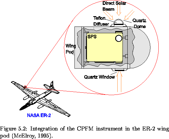
The CPFM instrument is a small, light-weight photodiode array spectrometer. It measures the solar flux on a horizontal surface, the vertical and horizontal polarized limb radiance components, and the along-track and cross-track polarized nadir radiance components. The limb observations consist of a ten-point scan. The detector is an EG&G 1024-element, photodiode array and is positioned at the focus of an f/2 concave, holographic diffraction grating. Filters are used to allow only light in either the first order, 375-775 nm, or the second order, 188-388 nm, to be focused onto the array. In addition, polarizers are used to select one polarization component of the limb or nadir radiance and reject the other. Due to the increased absorption by ozone combined with declining UV sensitivity there are no useful data below 300 nm. The layout of the CPFM spectroradiometer is shown in Figure 3.3. The CPFM instrument is installed in the ER-2 wing pod at the rear of the right wing, as shown in Figures 3.1 and 3.4.
The calibration for the absolute radiometry of the direct-viewing ports (limb and nadir fields) and the horizontal diffuser is carried out using a quartz-halogen lamp. The lamp calibration is accurate to 2% between 300 and 800 nm. Absolute radiance and irradiance were determined by scaling the measured signals by the signals observed when viewing the calibrated sources. The calibration process was carried out on-site as often as possible, including before the first flight and after the last. Wavelength calibration is done using emission lines from mercury and neon lamps. Each element of the array detector is associated with a wavelength using a cubic dispersion equation. Corrections are made post flight to account for wavelengths shifts caused by variation in pressure and temperature. The wavelength assignments are accurate to about 0.05 nm. Over the course of a flight, selected Neon and Fraunhoffer lines were observed to stray by a maximum of 0.1 nm. Stray light, or internally scattered light, becomes significant at wavelength less than 310 nm, contributing about 60% of the signal at 300 nm. Stray light is corrected for by subtracting off the signal between 290 and 295 nm, wavelengths at which there is no real light signal.
A single cycle of measurements consists of one complete spectrum (first and second order) for each of the five fields. The measurement sequence is horizontal flux, nadir (both polarizations) and limb (both polarizations). There is a 40-second delay while switching between the diffuser and nadir fields and a 30-second delay between the nadir and limb and between the limb and diffuser. The integration time used varied from 0.1 to 4 seconds.
The horizontal flux is measured by a transmitting diffuser which
approximates the performance of a cosine collector.
The angle between the diffuser normal and sun
(called the relative zenith angle) is measured and it is used to
cosine correct the measured flux.
Due to the surface properties of the diffuser, there is a departure
from true cosine behaviour and an additional correction must
be used.
For small relative zenith
angles this factor is small, about 2%, but increases to 20% at 85![]() .
For wavelengths greater than 500 nm, this correction factor is also
wavelength-dependent.
.
For wavelengths greater than 500 nm, this correction factor is also
wavelength-dependent.
The nadir and limb fields have a field of view of 0.1![]() by
10
by
10![]() .
In the nadir, with the ER-2 at 20 km, this results in
an observed surface area of 0.035 km
.
In the nadir, with the ER-2 at 20 km, this results in
an observed surface area of 0.035 km ![]() 3.5 km or approximately
0.1 km2. Switching between fields, as well as the different angles
in a limb scan, is accomplished by rotating a 45
3.5 km or approximately
0.1 km2. Switching between fields, as well as the different angles
in a limb scan, is accomplished by rotating a 45![]() prism using
a small motor.
To describe the look angle of the limb-viewing port, the elevation
angle is introduced. The elevation angle, EA, describes the angle
above (positive) or below (negative) the local horizon.
For any step in a scan, the elevation angle, EA, is given by the
following formula5.1,
prism using
a small motor.
To describe the look angle of the limb-viewing port, the elevation
angle is introduced. The elevation angle, EA, describes the angle
above (positive) or below (negative) the local horizon.
For any step in a scan, the elevation angle, EA, is given by the
following formula5.1,
| (5.1) |
The data used in this study will have passed through a number of processing steps. They are summarized below.
The slit function of the CPFM has been characterized by first finding
the peak intensity, normalizing to it, and determining
![]() .
This function has
three distinct regions, each of which is well approximated by
a linear fit. The slit function for the different regions are
(C.T. McElroy, personal communication, 1998),
.
This function has
three distinct regions, each of which is well approximated by
a linear fit. The slit function for the different regions are
(C.T. McElroy, personal communication, 1998),
![\begin{figure}% latex2html id marker 2145
\centering\leavevmode
\psfig{file=/hom...
...]
{Optical arrangement of CPFM spectroradiometer (McElroy, 1995).}
\end{figure}](img273.gif)
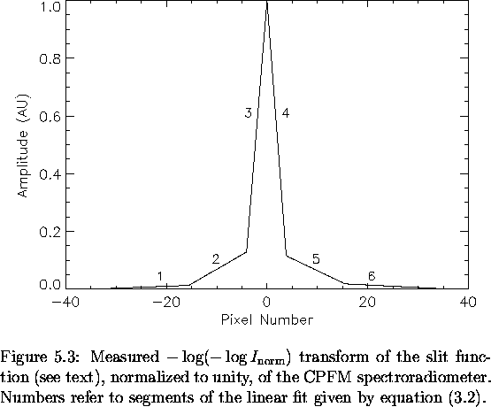
![\begin{displaymath}y = \left\{ \begin{array}{lcrclcc}
0.00456p + 0.1560 && -34.3...
...1 && 15.496 & \leq p \leq & 35.691 && [6]
\end{array} \right.
\end{displaymath}](img279.gif)